Our state of art technology suite provides timely diagnosis & better outcomes for your visual health.
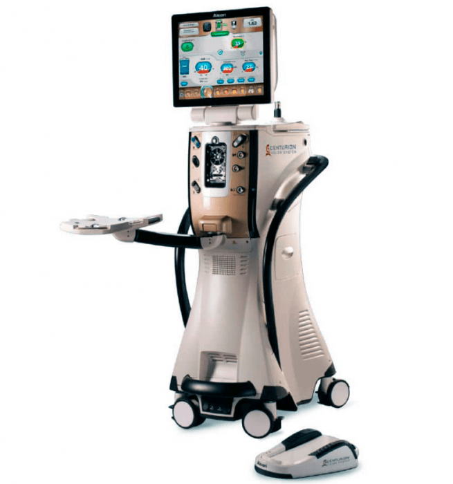
Centurion Vision System
It is designed to provide the surgeon with real-time, high-resolution video images of the eye during surgery, allowing for greater precision and control.
The system uses a digital camera and microscope to capture images of the eye, which are then displayed on a high-definition monitor. The surgeon can use a foot pedal to adjust the focus and zoom of the camera, as well as to switch between different lighting modes, such as bright field or red reflex.
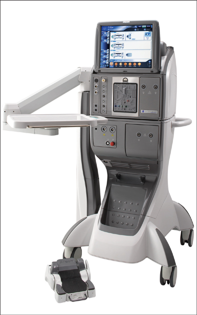
Constellation vision system
It is designed to provide the surgeon with high-quality visualization and precise control during delicate procedures in the back of the eye, such as those involving the retina, macula, and vitreous humor.
The system uses advanced imaging and illumination technologies, including a high-resolution digital camera and a range of lighting modes, to capture detailed images of the eye and display them on a high-definition monitor. The surgeon can use a foot pedal to adjust the focus, zoom, and other settings of the camera, as well as to switch between different illumination modes.
IOL master 700
The IOLMaster 700 is a non-invasive medical device used in ophthalmology to measure the length of the eye and other important parameters necessary for the selection and calculation of the intraocular lens (IOL) power in cataract surgery.
The IOLMaster 700 uses a technique called optical biometry, which uses light waves to measure the distance between the cornea and the retina and other important parameters of the eye. This method is highly accurate and provides more precise measurements than traditional ultrasound-based methods.
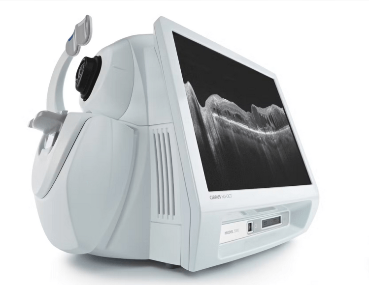
CIRRUS HD-OCT 500
The CIRRUS HD-OCT 500 is a high-resolution optical coherence tomography (OCT) imaging system used in ophthalmology to visualize and analyze the retina and optic nerve. It is a non-invasive diagnostic tool that provides detailed, cross-sectional images of the retina, allowing clinicians to detect and monitor a wide range of ocular diseases, including glaucoma, age-related macular degeneration, and diabetic retinopathy.
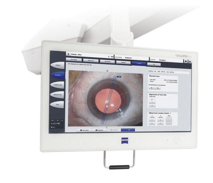
CALLISTO eye system
The CALLISTO eye system provides real-time image guidance during cataract surgery, enabling surgeons to achieve greater precision and accuracy. The system includes a high-resolution digital microscope, a 3D camera, and a tracking system that allows the surgeon to monitor the position of the patient’s eye and the surgical instruments in real time.
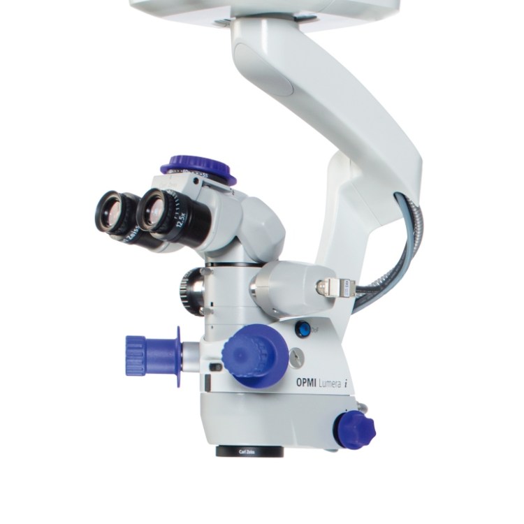
OPMI Lumera i microscope
The OPMI Lumera i microscope is designed to provide high-quality imaging during surgical procedures. It features an integrated camera system that captures high-resolution images, allowing the surgeon to visualize the surgical site in great detail. The microscope also has a range of illumination and visualization options, including fluorescence imaging and near-infrared imaging, which can help to enhance the contrast and clarity of the surgical field.
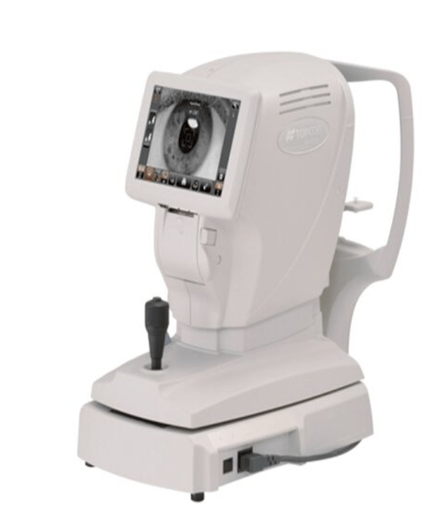
Auto Refractometer – Topcon
The Topcon Auto Refractometer is a diagnostic instrument used in optometry and ophthalmology to measure the refractive error of the eye. It is a non-invasive device that uses advanced technology to quickly and accurately measure the eye’s refractive error, which can help in the diagnosis of vision problems such as myopia, hyperopia, and astigmatism.
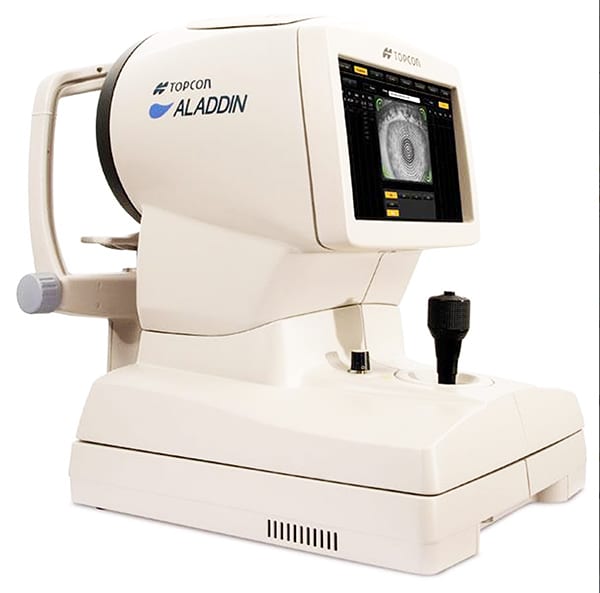
Topcon Optical Biometer with Typographer
Optical Biometer uses advanced technology to measure a range of parameters related to the eye, including axial length, corneal curvature, and anterior chamber depth. It also includes a topography function, which measures the shape and curvature of the cornea, allowing for a more comprehensive analysis of the eye’s refractive properties.
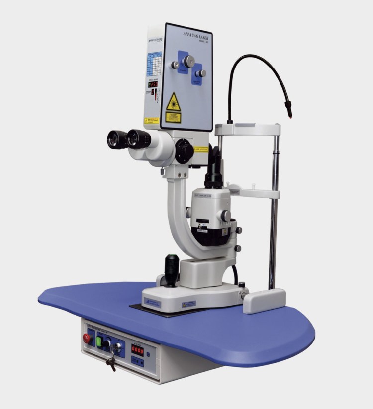
Yag Laser
A YAG laser, or yttrium aluminum garnet laser, is a type of medical laser used in ophthalmology for various procedures, including capsulotomy and iridotomy. It is a highly precise and focused laser that can create small, controlled incisions in the eye.
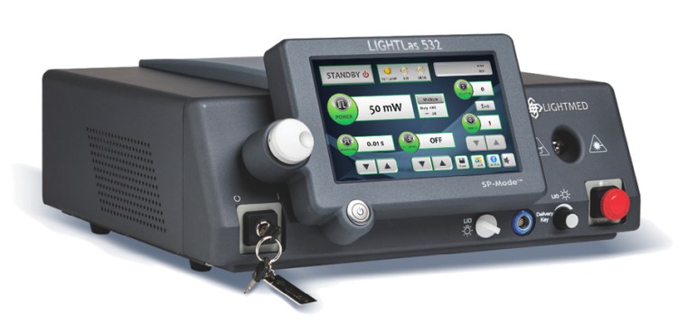
Green Laser 532 nm
A green laser with a wavelength of 532 nm is a type of medical laser used in ophthalmology for a range of procedures, including photocoagulation, trabeculoplasty, and capsulotomy. The green laser emits a highly focused beam of light that can be used to precisely target and treat specific areas of the eye.
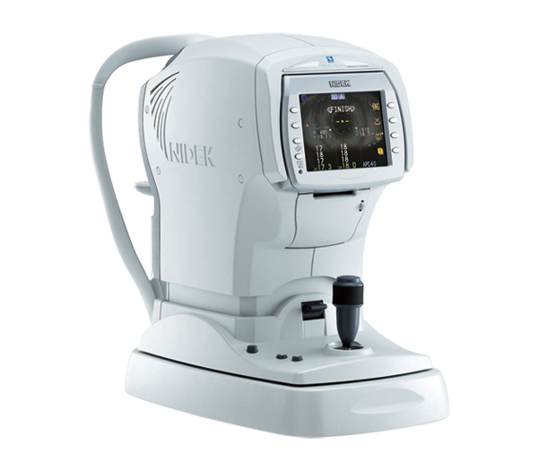
Nidek non-contact tonometer
The Nidek non-contact tonometer works by using a puff of air to measure IOP, rather than physically touching the eye like some other tonometers. The patient simply looks into the tonometer, and a quick puff of air is directed at the cornea. The tonometer then measures the pressure inside the eye based on how the cornea reacts to the puff of air.
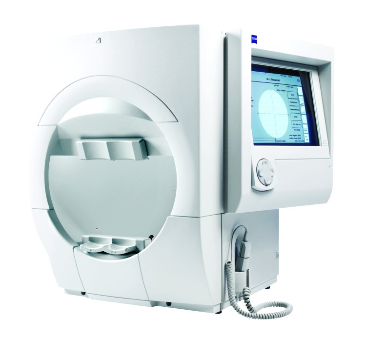
Humphrey visual field analyzer
The Humphrey Visual Field Analyzer works by projecting a series of light stimuli onto the patient’s retina while the patient focuses on a central point. The patient responds to the stimuli by pressing a button, and the analyzer maps the patient’s visual field based on their responses.
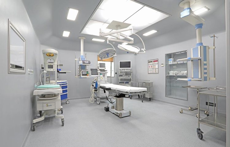
Modular operation theater
The modular construction process offers a number of benefits over traditional construction methods, including reduced construction time and cost, improved quality control, and greater flexibility in design and layout. Modular operating theaters can be designed to fit the specific needs of a hospital or healthcare facility, with customized layouts, equipment, and features that can help to improve patient outcomes and streamline surgical procedures.
Address
SA 15/171-D-1-5 Ghazipur Road Akatha Tiraha Opposite Rudra height, Paharia, Varanasi, Uttar Pradesh 221007
Timings
Our clinic is every day from 8:00 am to 2:00 pm, except the last month’s Sunday.
